The Value of Using Non-Invasive Technology for Level 3 Inspections
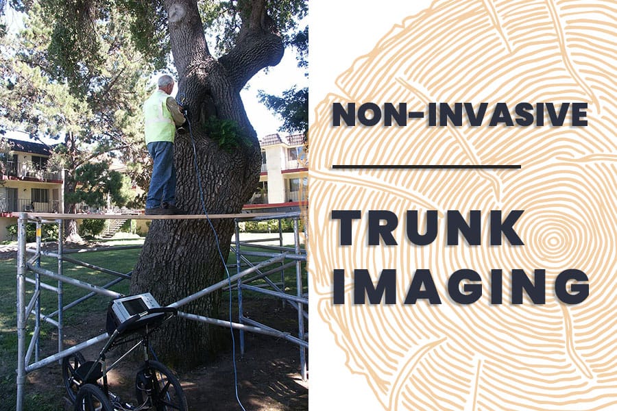
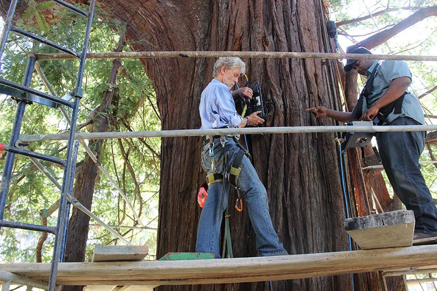
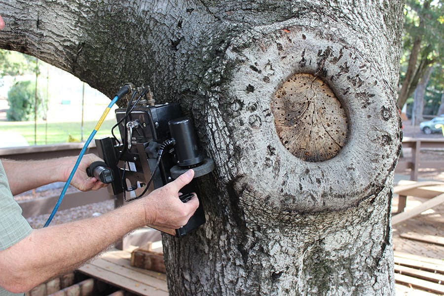
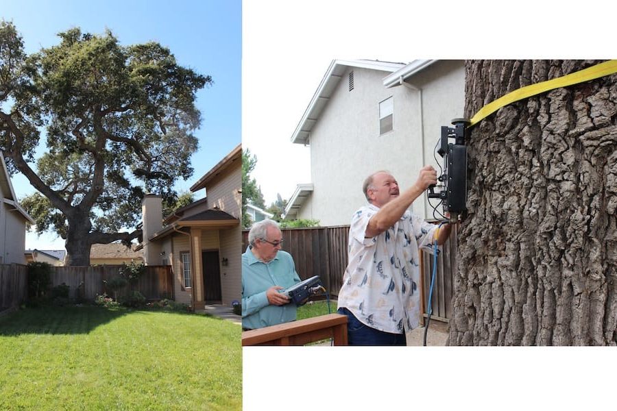
What benefit is there in using non-invasive diagnostic procedures during level 3 inspections? Trees can be adversely affected by the methods used to evaluate their health, particularly procedures that penetrate the bark.
The establishment of decay in living trees is affected by urban environmental stresses that range from a general weakening of the tree’s natural defense system to injuries that allow wood-rotting agents to gain entry through wounds. Trees have an internal protection system that uses a series of four internal walls, all beautifully designed to block the spread of disease causing pathogens within the tree. It’s referred to as CODIT (Compartmentalization of Decay in Trees).
When invasive testing methods are used, these protective walls can be pierced, allowing decay pathogens which at one time may have been localized or contained to spread within the tree.
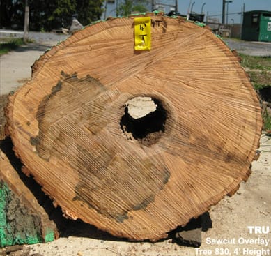
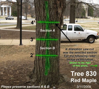
Case Study: Tree #830 – Acer rubrum
Location: University of Maryland Campus
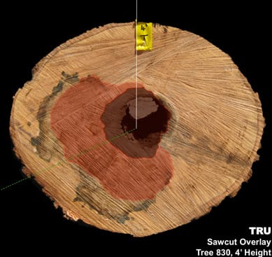
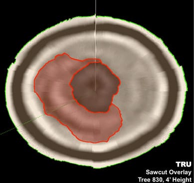
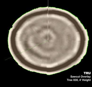
Specimen exhibited significant crown dieback and likely internal decay. It was designated for removal before TRU scanning. Excellent opportunity to compare actual and virtual (TRU) sawcuts.
*Images courtesy of Taylor Keene.

